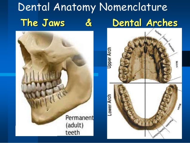
3D file Human Head Anatomy 3D printer design to downloadCults Biology Diagrams 1 Introduction to Dental Anatomy. Dental anatomy is defined here as, but is not limited to, the study of the development, morphology, function, and identity of each of the teeth in the human dentitions, as well as the way in which the teeth relate in shape, form, structure, color, and function to the other teeth in the same dental arch and to the teeth in the opposing arch. The dental arches are the two arches (crescent arrangements) of teeth, one on each jaw, that together constitute the dentition.In humans and many other species, the superior (maxillary or upper) dental arch is a little larger than the inferior (mandibular or lower) arch, so that in the normal condition the teeth in the maxilla (upper jaw) slightly overlap those of the mandible (lower jaw) both Dental anatomy is a field of anatomy dedicated to the study of human tooth structures. The development, appearance, and classification of teeth fall within its purview. For human teeth to have a healthy oral environment, enamel, dentin, It is located on the mandibular arch of the mouth, and generally opposes the maxillary first molars

This arched layout helps ensure a proper shape for your long-term dental health and a proper bite (with the upper teeth slightly in front of your lower teeth). You have two dental arch types, one upper (also called maxillary) and one lower (also called mandibular). The average adult has 32 permanent teeth, with 16 in their top arch and 16 in Dental Anatomy Learning Objectives. 1 Pronounce, define, and spell the Key Terms. 2 Explain the composition of each of the tissues of a tooth. 3 Use the correct terminology when describing the teeth. Dental Arches. In the human mouth, there are two dental arches: the maxillary and the mandibular.

1: Introduction to Dental Anatomy Biology Diagrams
Dental Arches. The oral cavity has two arches: the maxillary arch and the mandibular arch. The maxillary arch is part of the skull, is fixed, and incapable of movement. The teeth are set in the maxilla bone. The maxillary arch may be referred to as the "upper arch." The mandibular arch is moveable through the action of the temporomandibular In the adult permanent dentition, the mandibular dental arch (dentition of the mandibular dental arcade, lower dental arcade, or mandibular teeth) are the sixteen inferiorly located teeth that are embedded in the alveolar processes of the mandible. The mandibular dental arcade is equally divided into left and right quadrants. The teeth are aligned along the dental ridges and are surrounded by gingiva, forming the dental occlusion where the upper and lower teeth meet when the mouth is closed. Anatomy Each tooth is composed of multiple layers of tissues and is supported by surrounding structures like the periodontal ligament and gingiva. [1]

The human dentition is composed of two sets of teeth - primary and permanent.. Teeth are organised into two opposing arches - maxillary (upper) and mandibular (lower). These can be divided down the midline (mid-sagittal plane) into left and right halves. Teeth are positioned in alveolar sockets and connected to the bone by a suspensory periodontal ligament.

Oral Facial Anatomy Online Biology Diagrams
Atlas of dental anatomy: fully labeled illustrations of the teeth with dental terminology (orientation, surfaces, cusps, roots numbering systems) and detailed images of each permanent tooth Mandibular dental arch; Panoramic X-ray diagram; Deciduous teeth ; ANATOMICAL PARTS. Pocket Atlas of Human Anatomy: 5th edition - W. Dauber, Founded

