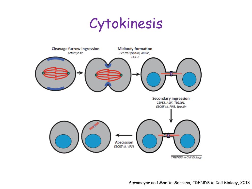
Rational engineering of new and improved cytokines Biology Diagrams Thus, many levels of RhoA regulation appear to be at work and generally feed through Ect2, MgcRacGAP, and F-actin. Cytokinesis completion depends on ESCRTIII-mediated lipid dynamics. Membrane trafficking and remodeling play significant roles at the abscission stage during cytokinesis. ESCRT is responsible for membrane constriction and fission. In a typical cell, cytokinesis accompanies every mitosis, although some cells, such as Drosophila embryos (discussed later) and vertebrate osteoclasts (discussed in Chapter 22), undergo mitosis without cytokinesis and become multinucleate. myosin II filaments, and many structural and regulatory proteins. Abstract. The primary goal of cytokinesis is to produce two daughter cells, each having a full set of chromosomes. To achieve this, cells assemble a dynamic structure between segregated sister chromatids called the contractile ring, which is made up of filamentous actin, myosin-II, and other regulatory proteins.
Second, endosomes may allow fast and localized delivery of regulatory proteins leading to the reorganization of the cytoskeleton and PM that is required for abscission to take place. In this mini-review we will not discuss the roles of specific lipids during cytokinesis, since several excellent reviews have been recently published on this topic The primary goal of cytokinesis is to produce two daughter cells, each having a full set of chromosomes. To achieve this, cells assemble a dynamic structure between segregated sister chromatids called the contractile ring, which is made up of filamentous actin, myosin-II, and other regulatory proteins. Constriction of the actomyosin ring generates a cleavage furrow that divides the cytoplasm

Classical and Emerging Regulatory Mechanisms of Cytokinesis in Animal ... Biology Diagrams
Mitosis and cytokinesis are key stages of cell division, each playing distinct roles in cellular replication. The regulation of these processes is tightly controlled by various molecular mechanisms. This article will explore the intricacies of mitosis, cytokinesis, and their regulation, highlighting the essential components and steps involved.
Many key cytokinesis proteins also perform important functions earlier in the cell cycle, especially during mitosis, and their removal leads to an arrest prior to cytokinesis. Using small molecules to study the regulation of cytokinesis. In addition to helping elucidate the critical role of actin and microtubules, small molecules have This ring is also stabilized by anchor proteins FtsA and ZipA, which tether FtsZ to the inner face of the plasma membrane. Z-ring also acts as a dynamic scaffold to recruit downstream proteins with critical regulatory functions of cytokinesis, such as peptidoglycan (PG) remodeling and chromosome segregation (den Blaauwen et al. 2017).

Membrane Dynamics during Cytokinesis Biology Diagrams
Cytokinesis, the final step of cell division, is a great example of robust cell shape regulation. A wide variety of cells ranging from the unicellular Dictyostelium to human cells in tissues proceed through highly similar, stereotypical cell shape changes during cell division. Typically, cells first round up forming a cleavage furrow in the middle, which constricts resulting in the formation