
Services Diagnostic Testing Biology Diagrams Understand the histology of human organs within the context of cell biology in preparation for further study of pathology, and advanced cell biology and molecular biology. Each Lecture covers a different system or region in detail. 0 LECTURES. 0 VIDEOS. Learning Outcomes. done. Histology Quizzes - Human Organ Systems. Complete over 1500 quiz questions to prepare for your formal histology examination. These extensive series of histology quiz questions are taken from content in "Meyer's Histology - Online Interactive Atlas" and 3 very well known Histology textbooks: Ovalle, W. K., Nahirney, P. C. and Netter, F. H. (2013

Atlas of Human Histology. A Guide to Microscopic Structure of Cells, Tissues and Organs. Robert L. Sorenson T. Clark Brelje. 3rd Edition Organ Systems. Organs are assembled from the four basic types of tissues and have cells with specialized functions. Chapter 9. Cardiovascular System. Chapter 10.

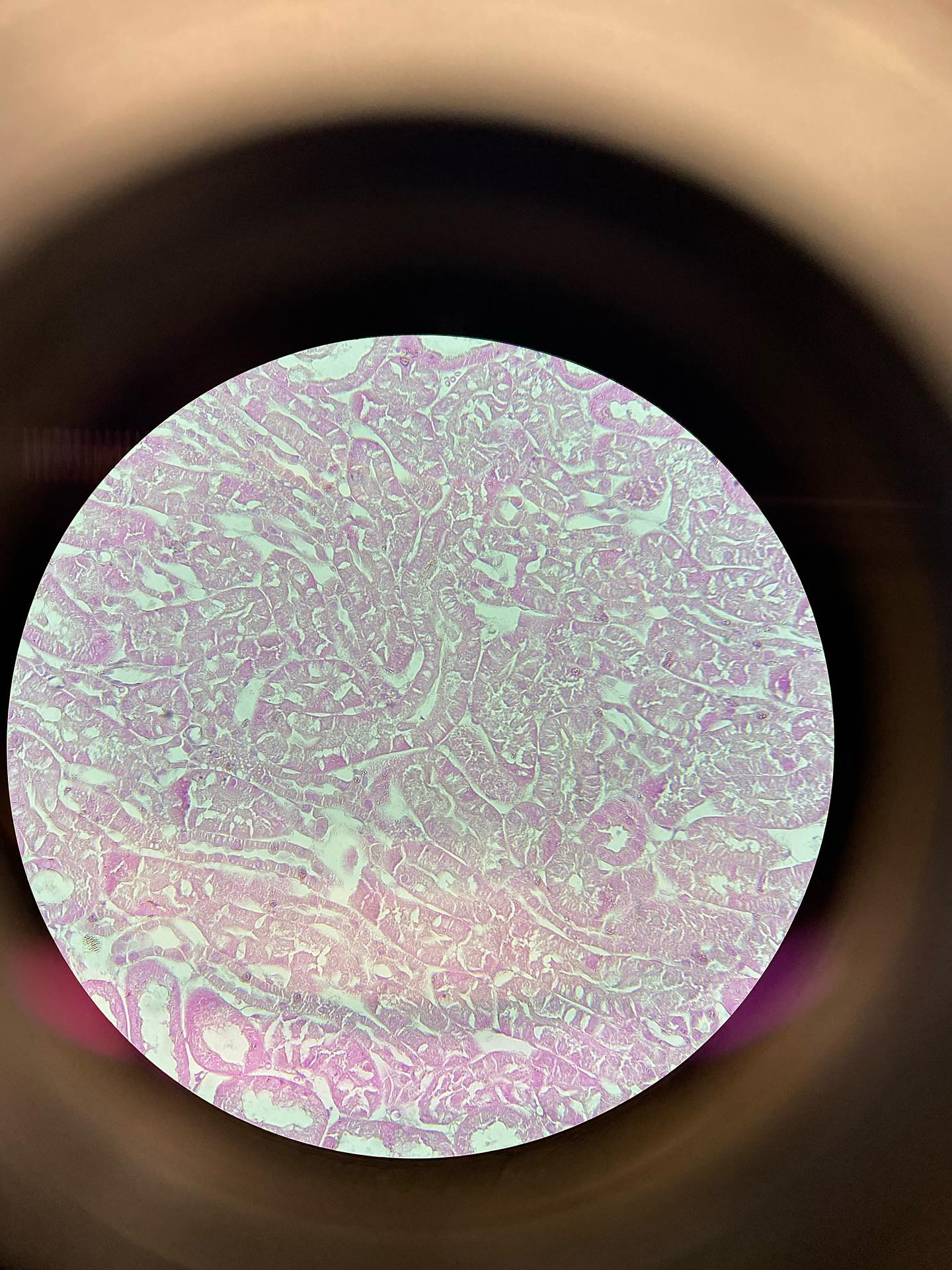
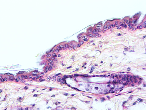
Histology of the Human Organs Biology Diagrams
Human anatomy is pretty straightforward. If you were to look at some bones on a skeleton, you'd see a greyish rigid mass with some bumps and depressions. However, if you take a much closer look, you'll see that the histology of bones, is a whole other story. Histology is the science of the microscopic structure of cells, tissues and organs Histology is the study of the microanatomy of cells, tissues, and organs as seen through a microscope. It examines the correlation between structure and function. The Atlas of Human Histology: A Guide to Microscopic Structure of Cells, Tissues and Organs by Robert L. Sorenson and T. Clark Brelje provides a print version of the core slides
Return to the Histology main menu. Histology Tutorials ; Basic histology is described, along with illustrative images, in this set of short tutorials arranged by organ system. Histology Correlations; A series of examples of abnormal histologic findings correlated with human disease conditions. Examples of Normal Histology.

Penn State Health Biology Diagrams
The Atlas of Human Histology: A Guide to Microscopic Structure of Cells, Tissues and Organs by Robert L. Sorenson and T. Clark Brelje is the companion atlas to the Histology Guide website providing a print version of the slides (1000 photomicrographs, 62 pages of text and 50 illustrations, 367 pages).
Atlas of Human Histology. A Guide to Microscopic Structure of Cells, Tissues and Organs. Robert L. Sorenson Organs. Organs are composed of more than one type of tissue snd its cells have specialized functions. Chapter 9 Histology Guide - a virtual histology laboratory with zoomable images of microscope slides and electron micrographs. histology is that it is the structural basis for cell, tissue and organ biology and function (physiology) and disease (pathology). What is the plan for the study of cells, tissues and organs? Histology is organized into four basic types of tissues. 1. Epithelium 2. Connective tissue including Cartilage and bone Blood and blood formation 3

(PDF) Atlas of Human Histology A Guide to Microscopic Structure of ... Biology Diagrams
Histology is the study of the tissues of the body and how these tissues are arranged to constitute organs. The Greek root histo can be translated as either "tissue" or "web" and both translations are appropriate because most tissues are webs of interwoven filaments and fibers, both cellular and noncellular, with membranous linings.
