
The Cell Cycle Biology Diagrams Anaphase chromosome separation in the acentrosomal meiosis I spindles of C. elegans oocytes is apparently independent of kinetochores . Instead, the chromosomes seem to be pushed from behind by microtubules growing and/or sliding out from the equator.
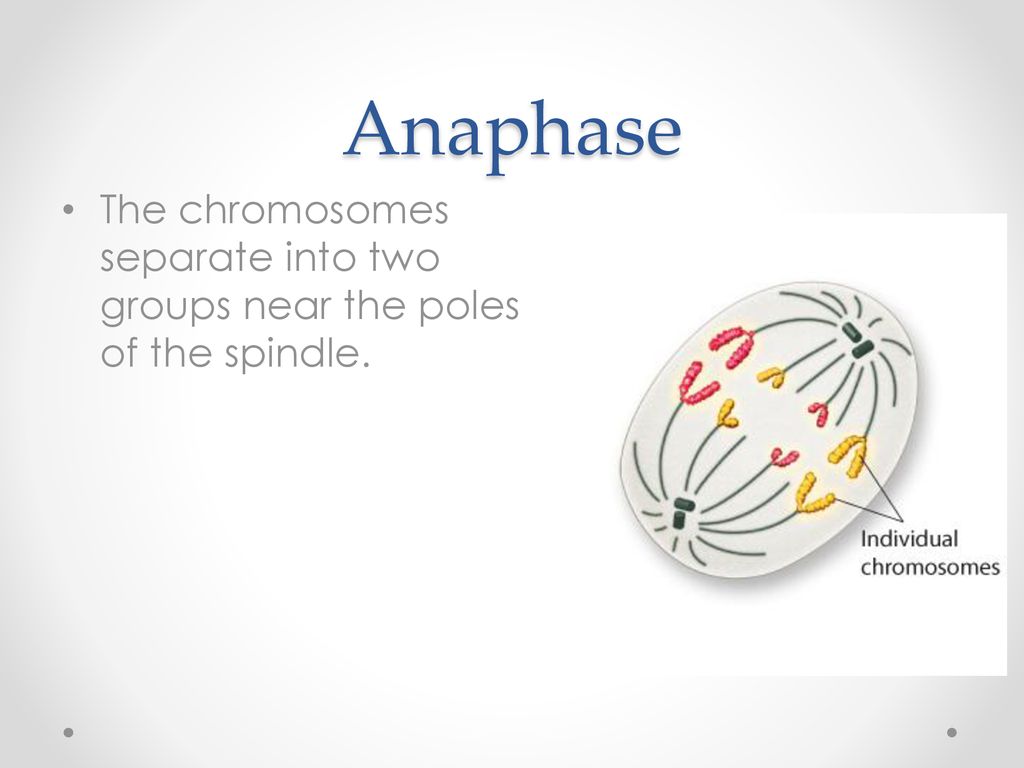
Separation occurs simultaneously at the centromere and each separated chromosome gets pulled by the spindles to the opposite poles of the cell. The function of anaphase is to ensure that each daughter cell receives identical sets of chromosomes before the final phase of the cell cycle, which is telophase.
Anaphase: The Separation of Chromatids Biology Diagrams
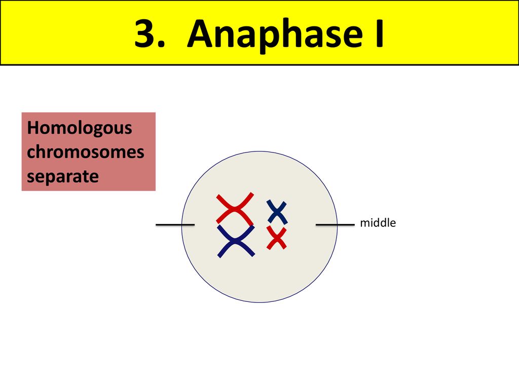
This mechanism separates homologous chromosomes into two separate groups. In anaphase I, the main goal of the spindle apparatus is apparent. As mentioned, a positive result to a spindle check must be completed before the move into anaphase, in which chromosome pairs are separated from their partners and transported to either side of the cell.
Anaphase: Chromatid Separation. Following chromosome alignment in metaphase, anaphase initiates the physical separation of sister chromatids. This stage involves cohesin breakdown, microtubule shortening, and chromatid movement toward opposite poles. Cohesin Cleavage
sister+chromatids+to+separate..jpg)
How Do Chromosomes Separate In Anaphase I? Biology Diagrams
Anaphase A involves the poleward movement of sister chromatids toward spindle poles, driven by the depolymerization of kinetochore microtubules and the activity of motor proteins, such as dynein and kinesin.Anaphase B is characterized by spindle elongation and separation of spindle poles, mediated by the sliding apart of antiparallel interpolar microtubules and the action of molecular motors The separation of chromosomes during Anaphase I is crucial for genetic diversity. It ensures that each gamete receives a unique combination of genes. This process, combined with crossing over in Prophase I and independent assortment, contributes significantly to variation among offspring. In contrast to Anaphase I, where homologous chromosomes separate, Anaphase II involves sister chromatids held together by cohesin proteins. The anaphase-promoting complex (APC) activates separase, an enzyme that cleaves cohesin, allowing chromatids to move to opposite poles. Spindle fibers exert tension on kinetochores, ensuring accurate

Anaphase Promoting Complex - The system of proteins, coenzymes, and other molecules that enable separase to degrade cohesin molecules, leading to separation of chromosomes. Cohesin - The protein molecules that bind sister chromatids or homologous chromosomes together.
